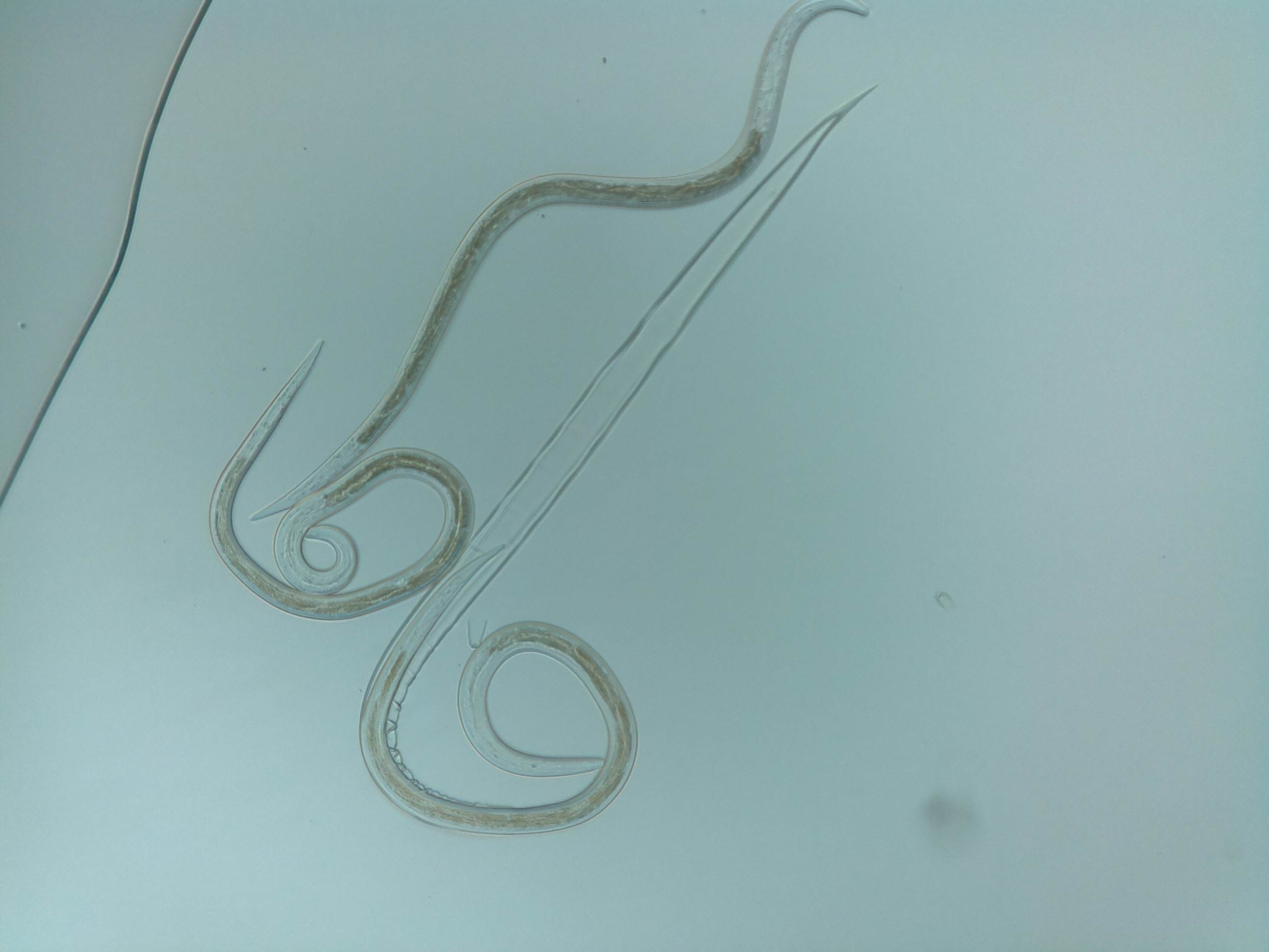
Sheep nematode worm shedding its sheath. Taken with the ioLight field microscope
All animals carry parasites, such as intestinal worms (or helminths). The parasites cause several diseases including gastritis and enteritis, which prevent the animal putting on weight. Some pathological parasites can even kill livestock.
The cost of parasites to farmers is large. The Animal Health drugs market is reported to be worth more than $7.5BN and many of these drugs are targeted on parasites. Companion animals, such as dogs and cats, are wormed regularly by their caring owners. However, parasites such as canine lungworm are killing increasing numbers of pets. This is why Bayer is using ioLight microscopes to promote Advocate®, which prevents infestation.
Governments worldwide are now introducing regulations to reduce the use of drugs in the food chain. Vets now have to diagnose intestinal parasites before treating farm animals. This is done by collecting a fecal sample from the back of the animal and sending it off to the lab for microscopic analysis to detect parasite eggs. This usually takes two days after which the vet returns to the farm to treat the animals if required. The ioLight portable field microscope is highly portable and has a wipe clean surface (which is important to vets working behind animals). It is also less expensive than a lab microscope and produces images on a tablet that can easily be shared with the vet’s clients, building that crucial trust between vet and farmer.
Moredun are one of the UK’s foremost labs working on animal parasites. They work with their member farmers and government to devise practical methods of controlling parasites. Moredun carry out standard faecal egg counts as described in a previous article; however they also study the life cycle of the parasites with the aim of increasing understanding to help to devise new methods of control.
Sheep nematodes have a fascinating life cycle – nematode eggs are laid inside sheep and leave the animal in the dung where they hatch into first stage larvae. They then shed their skin and become second stage larvae, remaining in the dung. Their skin is shed once more and they become third stage larvae at which point they leave the dung and move on to the grass where they are eaten by the sheep and the life cycle begins again.
Below is an image taken by Thomas Tzelos of Moredun using the ioLight field microscope. This image shows a sheep parasitic nematode, the brown stomach worm (aka Teladorsagia circumcincta, to give it it’s proper name!) The nematode is in the process of shedding its skin for the second time and becoming the infective third stage larvae.
The ioLight microscope provides a really convenient and easy to use way of imaging these larvae in the lab without all the complexity and cost of a conventional microscope.
The British government’s Centre for Environment, Fisheries and Aquaculture Science ( CEFAS ) has validated…
ioLight is delighted to welcome our latest Water Quality partner, Amore Aqua, based in Sweden.…
Recent press coverage has focused on water companies releasing sewage into rivers and lakes. How…
ioLight has developed a small, affordable and high-quality digital microscope for monitoring cell culture. The…
Counting stomata and observing their physiology tells us much about Plant growth and productivity. These…
ioLight announces successful funding round The world's leading compact microscope brand is delighted to announce…