ioLight has developed a small, affordable and high-quality digital microscope for monitoring cell culture. The product is fully sealed so it can be left inside the chamber and easily sterilised afterwards. It sends images wirelessly to a tablet or computer outside the incubator so that the cell culture can be viewed without opening the incubator and risking damaging the culture.
Having a microscope in the incubator allows for continuous monitoring of cultures, giving insights into cell culture health and early detection of contaminants. This improves yields and allows experiments to be carried out directly in the incubator, providing a stable and contaminant-free environment for long-running assays or delicate 3D cultures.
The compact size of the ioLight inverted microscope also frees up space. Whether that is for other flasks containing the rest of the current batch or the next experiment. The instrument is robust and sealed, so it can operate in the high humidity and CO2 conditions in an incubator and be fully sterilised to prevent contamination after each run.
Furthermore, the ioLight cell culture microscope transmits images wirelessly, so the microscope can simply be placed into an incubator to provide continuous monitoring with timelapse and video capture features. Your cell cultures can be presented in standard Petri dishes, 96 well plates or T-flasks. The microscope has settings for brightfield, Low NA (high contrast), digital phase contrast and darkfield to provide good images even in low-contrast subjects.
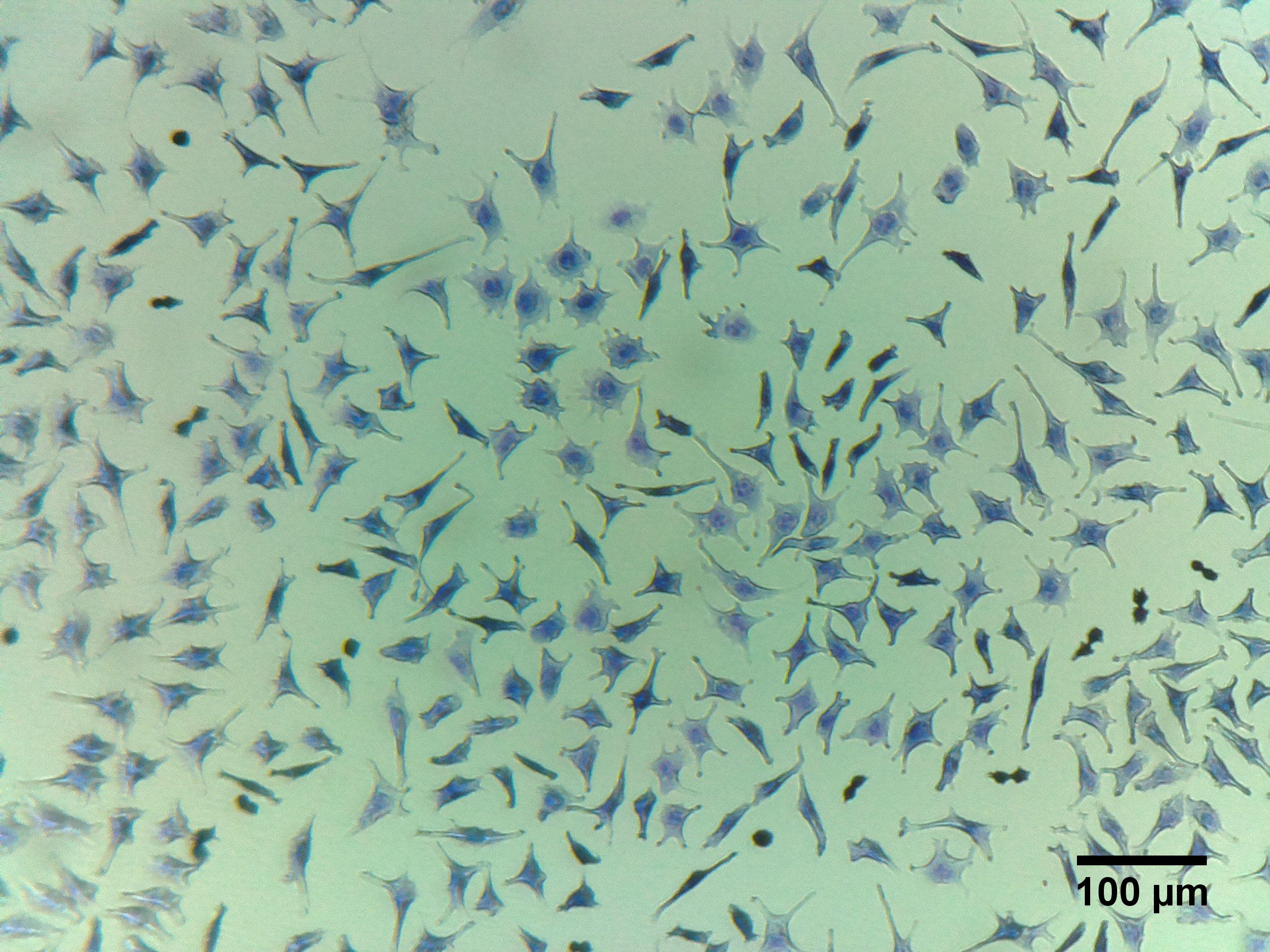
The image and YouTube video were taken inside an incubator. They clearly show the number, motility, confluence and morphology of the cells, providing researchers and technicians with confidence in the health of their experimental cell lines. Our thanks to Dr Mark Willet at The University of Southampton for helping us produce these images.
ioLight is pleased to announce that we are working with Ramboll Group and Lake Associations in Wisconsin and New York State to identify the toxic algae that contaminate lakes, rivers and oceans worldwide.
Many algal blooms are in fact harmless, but a safe diagnosis requires a microscope and an expert. Lake Association members often drive samples long distances to the Lab to have their samples checked so that their lake can be reopened. It can be several days to confirm that the lake is safe.
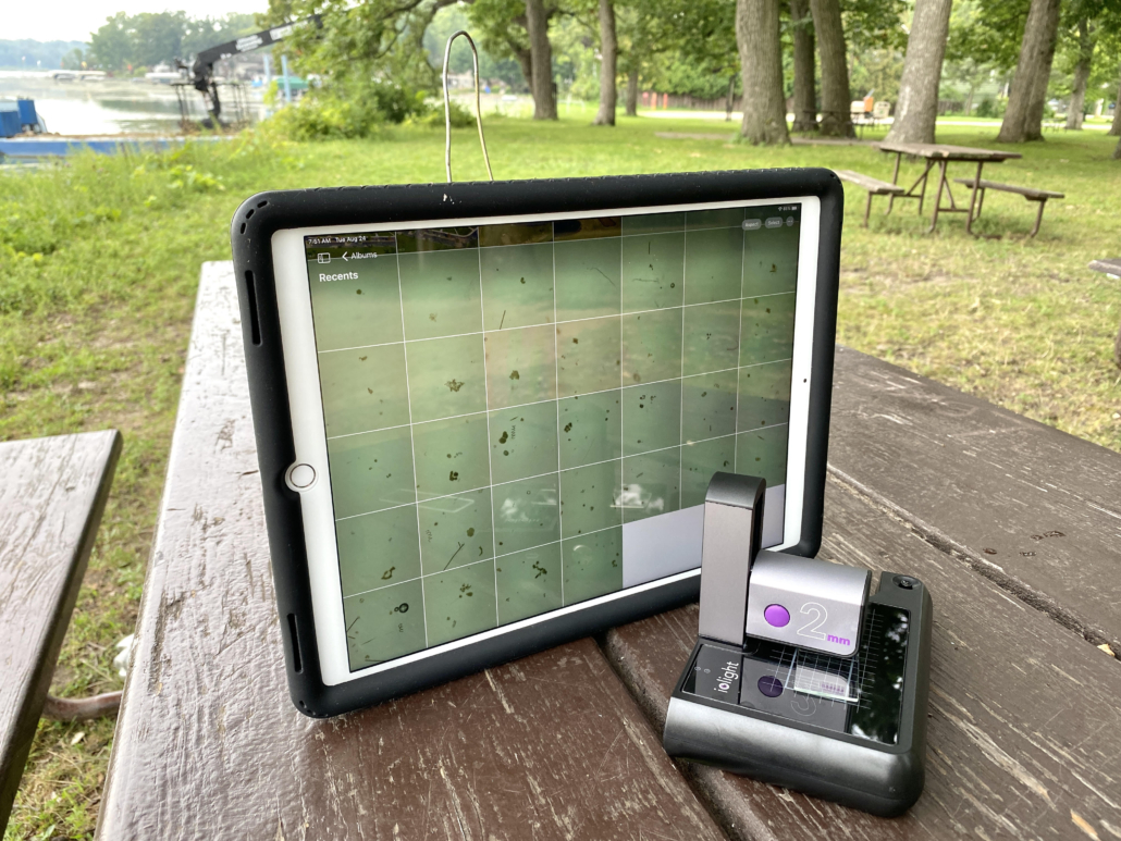
Ramboll spinout, BloomOptix is developing an Artificial Intelligence app to give an instant result on a mobile phone. The expert app is currently being trained using images from lake associations in Wisconsin and New York State. Samples are collected from the lake and placed on the platform of an ioLight pocket microscope and saved onto a mobile phone for analysis. The app will cut the time to diagnose Harmful Algal Blooms from days to minutes. This will maximise the time we can enjoy our beaches and reduce losses to businesses that rely on tourism.
Andrew Monk, CEO of ioLight says “the ioLight microscope is compact and simple for anyone to use, yet still produces high enough quality images for the BloomOptix app to identify toxic algae”. Bryan Arndt, Managing Consultant at Ramboll Water says “The ioLight microscope is twice the quality and half the price of other microscopes we tested.”
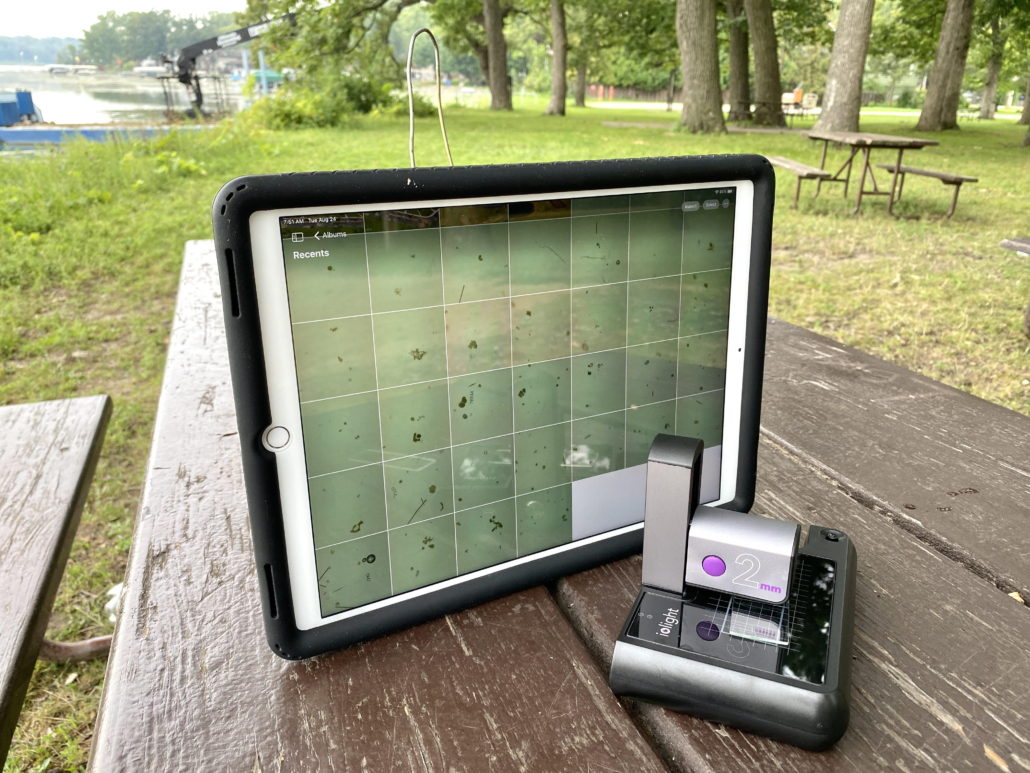
ioLight microscope counting algae on a lakeside in the Yahara River watershed, Wisconsin. Image courtesy of Bob Bertra Clean Lake Alliance.
About Ramboll
Ramboll is a global engineering, architecture and consultancy company founded in Denmark in 1945.
Their 16,000 experts create sustainable solutions across Buildings; Transport; Water; Environment & Health; Architecture, Landscape & Urbanism; Energy and Management Consulting.
Across the world, Ramboll combines local experience with a global knowledge base to create sustainable cities and societies. They combine insights with the power to drive positive change to our clients, in the form of ideas that can be realised and implemented.
ioLight has patented the only pocket microscope that can share images of single-cell organisms such as algae and diatoms. ioLight’s solutions allow professional and amateur scientists to measure the effects of climate change such as Harmful Algal Blooms.
ioLight’s customers have taken their microscopes to The Arctic, Antarctic, The Amazon and even up Mount Everest where conventional microscopes cannot be used.
ioLight’s Magnificent Mobile Microscope goes wherever researchers go.
Cells grown in labs are a mainstay of biomedical research. Scientists have available to them a wide variety of carefully selected cells, cultivated from animals and humans. These cells can be grown in the lab so that scientists can test potential therapies, drugs and vaccines on appropriately selected cells to see the effect and then infer what might be the effect when used on live humans and animals.
A cell line is created by harvesting some cells from an animal or human and growing them in flasks. As the cells grow they multiply, and so by continually cultivating the cells, there is a constant supply of new cells identical to the original cells.
There are a large variety of cell lines for various parts of many animals. Each is suitable for different types of experiments and for investigating different biological processes. For example, the CHO-K1 cell line is epithelial cells that come from a particular Chinese Hamster’s Ovary (hence CHO) and is commonly used to test protein therapeutics. Calu-3 is another example of a cell line – this was originally taken from an individual human lung cancer, and so when it is used in the lab, it is often used to investigate and test cancer therapies.
Cells are widely used across a variety of biomedical research topics, but regardless of the research topic, the cell culture techniques required are very similar. A whole industry has grown up to provide cell culture techniques and equipment.
Cells need very carefully controlled conditions to grow successfully, to provide these conditions an incubator is used. The incubator promotes cell growth by maintaining the optimum temperature, humidity, and often atmosphere. Cells typically take between many hours and days to grow, and for the effects of the experiments to be visible. Thus the cells need to be imaged regularly over the course of hours/days both to determine a) that the cells are growing as expected and b) to see the effect of the experiment on the cells. Conventionally this is done by removing the flask from the incubator, putting it on a microscope and looking at the cells. However moving the flask and taking it out of the controlled incubator environment adversely affects the cells, so images are recorded infrequently to minimise the disruption to the cells.
What is Live Cell Imaging?
Live Cell Imaging refers to recording microscope images of the cells without moving the cell culture flask or removing it from the incubator. This minimises failed experiments and enables the scientist to better track the effect of the experiment on the cells. This improves the quality of the data and gives a better understanding of the experiment.
Conventional microscopes are large, complex and cannot be used inside incubators. Instead, micro-incubator chambers that fit on microscope stages are used. However, this approach is expensive and ties up a valuable bench microscope for the entire duration of the cell culture, which makes this approach impractical for most research projects.
Recently, miniature digital inverted microscopes have become available. These microscopes image cell flasks, dishes or well plates from below and record images whenever the researcher wants without disturbing the cells. They are small enough to fit inside standard incubators, making Live Cell Imaging accessible to a much wider range of budgets.
Many of these inverted incubator microscopes are supplied with sophisticated software to automatically track cell development and provide the researcher with high-level data on the progress of cell growth. This is appropriate for many large scale development labs. However, much cutting-edge research is done by the smaller labs for whom this level of sophistication and cost is not appropriate.
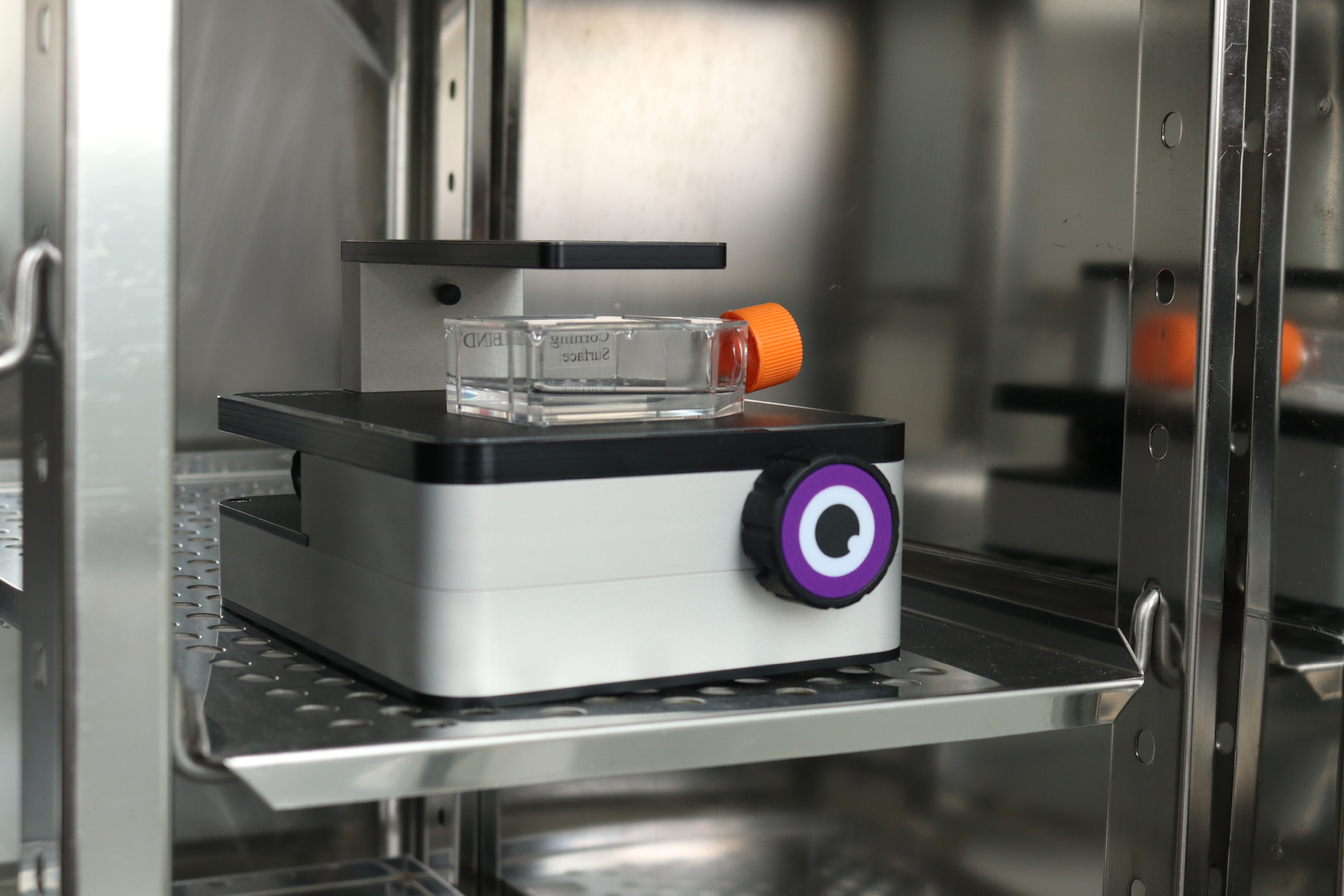
Above: Inverted digital microscope inside an incubator
The images below are taken using a simple low-cost inverted incubator microscope made by ioLight (iolight.co.uk). This microscope fits inside standard incubators and sends images via WiFi to a tablet or computer in the lab. It can record time-lapse images which can be viewed remotely, so the researcher can monitor cells for days without going into the lab or removing the culture from the incubator. This reduces the number of failed cell cultures and provides better data on the results of the experiments.
This type of affordable digital inverted microscope is suitable for imaging a wide variety of cells as well as a myriad of other samples. Some example images of cells are shown below:
Live Cell Imaging: Hepatocytes
Hepatocyte cells make up the majority of the cells of a liver and are involved in a range of functions including protein synthesis and storage and transformation of carbohydrates and formation of bile.
Fibroblasts are cells that synthesize the structural framework for animal tissues. They are the most common cells of animal connective tissue. They are used in a variety of applications including stem cell research, and vaccine development.
These cells are cloned from bone marrow and are particular types of neuronal cells often used to study Parkinson’s disease, neurogenesis and other characteristics of brain cells.
Live Cell Imaging is the best way of ensuring a cell culture is progressing correctly and reduces the time and money wasted on failed cell cultures. Live Cell Imaging also enables the researcher to record better data on the effect of the experiment on the cells, thus improving the quality of research. Until recently Live Cell Imaging has been very expensive and the preserve of large labs. However, simpler Live Cell Imaging devices such as the ioLight inverted microscope are now available. Smaller labs, which are often where much of the cutting edge research is done, can use these new microscopes to access many of the benefits of more costly Live Cell Imagers.
https://iolight.co.uk/wp-content/uploads/2021/09/IMG_8614-scaled.jpg13332000ioLighthttp://iolight.co.uk/wp-content/uploads/2016/01/iolight-logo.pngioLight2021-09-02 16:08:192021-09-06 14:35:58Live Cell Imaging on a budget
Last week ioLight completed our second webinar training citizen scientists in the USA to use the ioLight microscope to track Harmful Algae Blooms.
The State of New York (NY) has approximately 8000 lakes, managed by 200 Lake Associations. These are made up of residents living on the shores of the lake who have a strong interest in maintaining the health of their lake. Suspect algal blooms are reported to the NY Department of Environmental Conservation, which may issue an advisory notice closing the lake to recreational activities.
Professor Greg Boyer of the State University of New York (SUNY) College of Environmental Science and Forestry (ESF) says that many of these blooms are in fact harmless, but diagnosis requires a microscope for visual identification. Lake Association members often drive long distances to bring samples to the lab in Syracuse NY to have their samples checked to ensure their lake is safe.
We have now trained a team of 16 Citizen Scientists who are taking images of algae to train an AI mobile phone app to differentiate between toxic and harmless species. One of the Clean Lake Alliance volunteers reported “I just wanted to say what a great device it is. I take it with me to the lake and capture wonderful images of lake algae. The link to the iPad works like a charm.” He also captured the picture below.
ioLight trains citizen scientists to teach the microscope to track harmful algae
Once the AI has analysed large numbers of algae images it will be trained to deploy more widely for the next algae season in the summer of 2022. We anticipate delivering up to 300 ioLight microscopes to lakes across North America next year. This valuable work will protect visitors to the lakes and keep beaches open for leisure activities through the summer.
https://iolight.co.uk/wp-content/uploads/2021/08/bobertera-scaled-e1630419157980.jpg7501000ioLighthttp://iolight.co.uk/wp-content/uploads/2016/01/iolight-logo.pngioLight2021-08-31 15:09:142024-10-24 11:13:33US Citizen Scientists teach AI to recognise Harmful Algae Blooms
ioLight Magnificent Mobile Microscopes is excited to announce a new partnership with School Finance Direct to help UK schools to acquire the latest robust high-resolution portable microscopes.
ioLight’s portable high-resolution microscopes are now available with finance from School Finance Direct
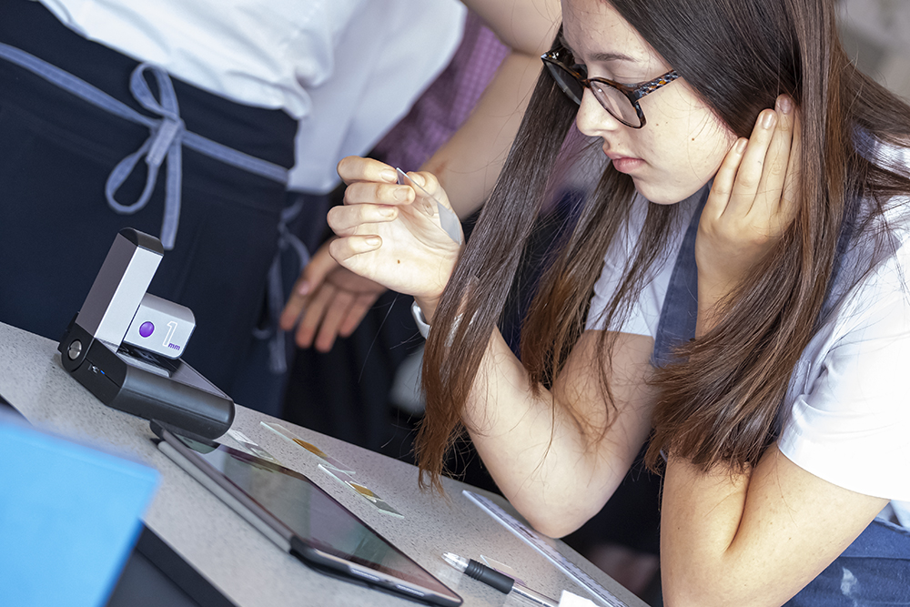
ioLight microscopes are wonderful teaching tools. They are simple to use for scientists as young as 8 and display beautiful images of protozoa, algae and cells on any mobile phone, ready for sharing and pasting into projects. Young people find mobile phone technology easier to use than traditional optics and ioLight’s products open a window to the building blocks of life using technology familiar to all young people.
1-micron resolution images can be displayed on classroom whiteboards for discussion with the wider group and the robust device can be thrown into a bag for field trips. Dr Richard Wilson, Head of Science at Testbourne Community School says “The ioLight microscopes are brilliant communication tools for practical science. Students can easily use them to see clear living images on a big screen anywhere, without squinting through eyepieces.” Dr Wilson continues “Students have particularly enjoyed being able to project live feeds from the ioLight microscope onto the classroom board to share their work with their peers and I’ve valued the flexibility as Head of Department of being able to use the ioLight microscope with any of our department laptops, teacher desktops, and our iPads and Android tablets.”
Schools Finance Direct was established in response to the Government’s Department for Education (DfE) guidance for schools publications and overwhelming demand from those organising finance for schools for practical real-world solutions. Adrian Langford, CEO of School Finance Direct says “Throughout our extensive and ongoing research, we recognised:
The education sector deserves a better deal
Those in schools responsible for organising resources deserve a better deal
People trying to make a difference need the right resources to get the job done”.
Langford adds “As a direct funder to the education sector, our objective is to exceed the nationwide requirement for those organising finance for schools to have direct access to finance facilities that provide value, transparency, compliance and support with levels of service specifically designed to smooth the whole process.”
Andrew Monk CEO of ioLight says “school science departments tell us how useful the ioLight microscope is for inspiring young scientists. Our partnership with School Finance Direct now makes this valuable teaching tool available to all schools.”
https://iolight.co.uk/wp-content/uploads/2021/08/testbourne-0156.jpg6671000ioLighthttp://iolight.co.uk/wp-content/uploads/2016/01/iolight-logo.pngioLight2021-08-25 10:22:332021-08-25 10:27:31ioLight partners with School Finance Direct
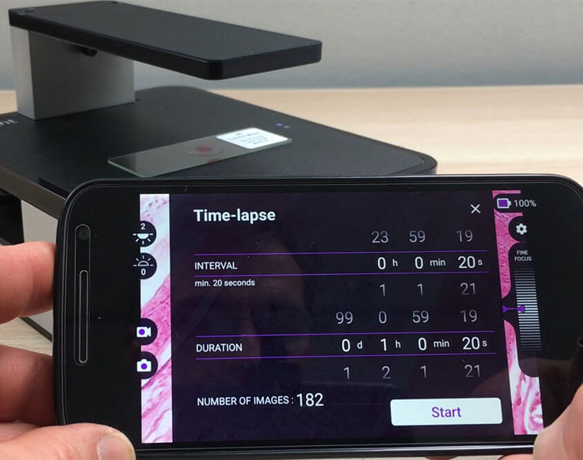
ioLight is pleased to announce the beta release of its time-lapse Android app. The app has all of the features of the standard Android app, with live video recording, beautiful 1 micron resolution images and wireless focus. Now, the new Android App beta release also includes programmable time-lapse recording to capture images at fixed intervals from 20 seconds up to 24 hours and run times up to 99 days.
The user interface is simple and flexible, it even reminds you to check that there is space on your device for the images to be stored, ready to upload to Google Photos or your favourite image-sharing platform. Once the images are uploaded, they can be viewed from anywhere on the internet at any time without going into the lab (this requires the ioLight Internet Module iIM).
The time-lapse Android app works well with any ioLight microscope, but it is ideal for the inverted microscope. This product is a small footprint cell scanner that can be mounted inside a CO2 incubator or bio-isolator and transmits images to the Android device outside. This means there is no need to open the door and risk contamination or disruption of the experimental conditions. The inverted microscope is sealed against the damp humid atmospheres of incubators and VHP cleaning and can view cultures in most Petri dishes, well plates and flasks as well as on slides.
The combination of the inverted microscope, timelapse app and ioLight Internet Module provides a compact, cost-effective platform for remote viewing of cell cultures 24/7. Find out how to get your time-lapse app for Android and ioLight Internet Module from info@iolight.co.uk.
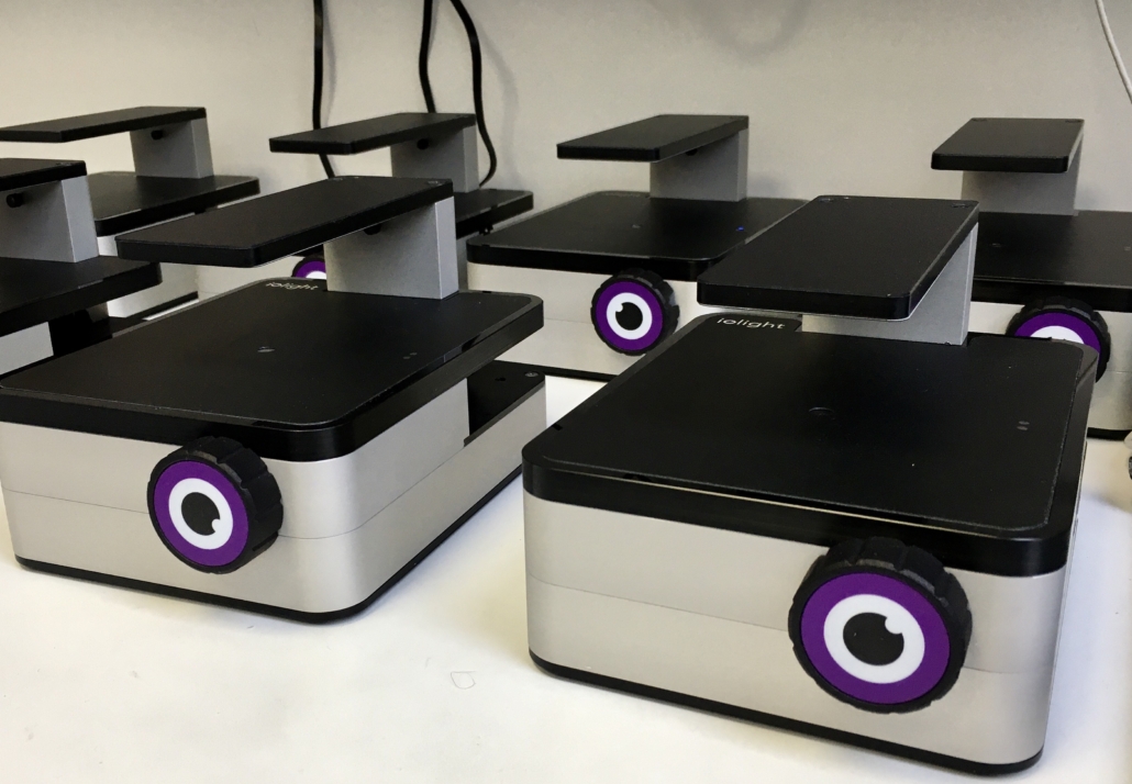
ioLight’s new inverted microscope is now available ex stock. The compact laboratory product is small enough to fit inside a CO2 incubator and is now available ex stock from ioLight’s webstore.
ioLight’s new inverted microscope uses the company’s robust platform which has been used in the field in Antarctica, the Amazon Rainforest and even on Mount Everest. The new inverted microscope wraps the microscope in a sealed package for use inside incubators in biological laboratories. The sealed case is resistant to the damp acidic atmospheres and it means that researchers can monitor their experiments without opening the door or disrupting their experiment. Andrew Monk, CEO of ioLight says “the ability to use a microscope to view slides, Petri dishes, well plates or flasks inside the incubator is a real benefit, as opening the door risks invalidating the results”, he adds that “We are working on an additional feature that could allow researchers to monitor experiments over the internet from home. No more trips to the lab to monitor progress in the middle of the night!”
The new inverted microscope has an improved resolution of 1 micron and even better image quality. This makes it the perfect tool to monitor cell development and tissue growth inside incubators and other small laboratory experiments. It still works outside too, with the included battery providing all-day power to view algae, protozoa and seaweed at the beach or lakeshore. Beautiful ioLight images are displayed directly to your cellphone or tablet screen for instant sharing.
https://iolight.co.uk/wp-content/uploads/2021/02/IMG_E2193-scaled.jpg13872000ioLighthttp://iolight.co.uk/wp-content/uploads/2016/01/iolight-logo.pngioLight2021-02-22 18:04:202021-02-22 18:11:18New inverted microscope now available
ioLight is pleased to announce the launch of our new inverted microscope for use inside incubators.
The CO2 incubator is used in most bio labs to grow cells, tissues and embryos. CO2 is used to exclude oxygen and the temperature is controlled at 37°C or 98°F to emulate in vivo conditions. The new ioLight product is sealed against this atmosphere and can be left inside the incubator for the duration of the experiment. ioLight’s wireless connection allows researchers to monitor the progress of their experiment without opening the chamber door and disturbing experimental conditions, reducing experimental errors.
Live cell growth view remote
The new release of ioLight’s inverted microscope features a new top illuminator with normal bright field, low NA and dark field illumination modes programmable from the free app or web browser. The height of the illuminator has been increased to accommodate 40mm flasks and well plates as well as Petri dishes and slides. The new glass optical system delivers the same crystal clear 1 micron resolution as our field microscopes.
ioLight’s folding field microscopes have been used in the Amazon Rainforest, Antarctica and even on Mount Everest. The same high resolution, compact and robust designs are used for the inverted microscope. ioLight’s CEO Andrew Monk says “we are really excited about this microscope, which deploys the world’s toughest portable microscope in laboratories too.” He continues “space is often at a premium inside laboratory experiments and not many microscopes are tough enough to survive the warm, humid and acidic atmosphere inside a CO2 incubator. This microscope brings ioLight out of the jungles, mountains and frozen lands where we normally work and into the laboratory.”
The ioLight inverted microscope saves its images onto your tablet or computer in standard file formats that can be shared easily and analysed using your favourite generic image processing software. True to its ioLight roots, the inverted microscope still has a battery so it is perfect for marine biologists to use at the water’s edge too.
https://iolight.co.uk/wp-content/uploads/2021/01/IMG_E2039-scaled.jpg13292000ioLighthttp://iolight.co.uk/wp-content/uploads/2016/01/iolight-logo.pngioLight2021-01-25 18:00:482021-01-25 18:06:42New microscope works inside laboratory incubators
Biology teachers at colleges and universities around the world are carefully working out how to teach in a post-virus world. A lot of work has been invested in planning teaching scenarios from further lockdowns to a return to the ‘new normal’. Video conferencing and teaching platforms such as Zoom, Classroom and Teams are already a daily part of teaching and learning.
Biology is one of the more difficult subjects in which to plan a new normal. The best teachers inspire their students, showing them samples of organisms at macroscopic and microscopic levels. This encourages their audience to connect biological processes all the way from global ecology to the biochemistry of a cell. The microscope is the key tool in this journey because it connects the global environment to cell biology. With most teaching laboratories closed during lockdown and field trips cancelled, inspirational teaching of biology has become really challenging. Biology teachers are intrinsically creative and many are looking for microscopes that their students could use when the teaching labs are closed.
Microscopes for $20?
Just $20 buys a contact lens that clips onto the camera of a student’s mobile phone. As a general-purpose magnifier, these are great for low magnification applications such as plant structure. With care, they can work in applications that need more magnification over a small area. However, the field of view of clip-on lenses is limited and images are usually surrounded by a black circle and out of focus at the edges. Nevertheless, this is a useful and inexpensive introduction to small animals and plants.
For a slightly larger investment, schools and colleges can purchase a digital magnifier that looks like a flashlight and connects to a computer with a USB cable or WiFi. These devices are either handheld or stand-mounted to give more stability. They often claim x1000 magnification, but in practice, the resolution is typically more like 50 microns – the thickness of a human hair. Which makes them pretty useful for electronics and insect anatomy.
Teaching biology over video
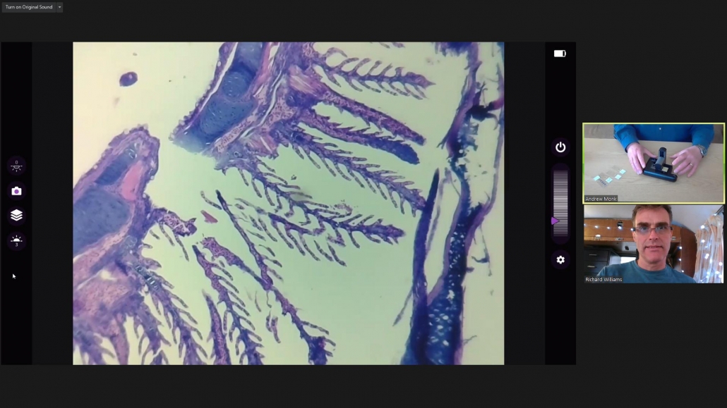
ioLight microscope displaying a high-resolution image on a Zoom conference call
Teaching cell structure needs a resolution of 1 micron and in the past, this was only available from a laboratory compound microscope. UK startup ioLight Ltd has patented a portable microscope with a stage and transmitted illumination. This pocket-sized instrument delivers 1 micron resolution images to any smartphone or tablet ready for sharing or pasting into projects. The performance of the ioLight product is similar to that of a lab microscope with two important differences: it is portable and internet-connected.
The robust ioLight microscope fits in a jacket pocket and can easily be posted to locked-down students for socially distanced field trips at home. The powerful 1 micron resolution allows students to image animal and plant cells, protozoa and algae. In fact, anything that a college laboratory microscope can see. Its portable format makes it perfect for citizen science surveys such as Microplastics or Harmful Algal Blooms (with a proper risk assessment).
The internet connectivity of the ioLight microscope means that video platforms such as Zoom, Classroom and Teams can use it to teach microscopy online, allowing students to see clearly the instructor’s microscope screen. This exciting development allows teachers to deliver microscopy classes online and remotely. ioLight even offers a fluorescence version of its product, which allows colleges and universities to introduce students to one of the central tools of microbiology.
Who knows, perhaps your students could even tag the COVID-19 virus and find a cure.
https://iolight.co.uk/wp-content/uploads/2020/07/Clipboard09.jpg10781917ioLighthttp://iolight.co.uk/wp-content/uploads/2016/01/iolight-logo.pngioLight2020-07-16 16:22:122020-08-14 14:05:17Biology teaching in a post virus class
National Geographic monitors the impact of climate change in Nepal
Climate Change is driving life in the Himalayas to move up the mountains as the lower altitudes warm. The latest National Geographic video documents Tracie Seimon leading a team of biologists carrying out a biodiversity study to monitor this movement. Far from her laboratory at the Wildlife Conservation Society’s Bronx Zoo in New York, Tracie and the team of local scientists use the ioLight microscope to monitor protozoa at high altitude. The ioLight microscope weighs just one pound and records high-resolution images and videos on a standard mobile phone. The microscope is extremely rugged and weighs just one pound, so it easily fits into a rucksack pocket.
Biology students use the ioLight microscope in the Amazon
University biology departments use ioLight in post COVID-19 teaching
Closer to home university biology departments are working out how to research and teach climate change in the wake of COVID-19. The ioLight microscope allows students to work in any environment in small physically distanced groups. Working away from the laboratory minimises the risk of spreading the virus and gives students a real first-hand experience of the environment.
https://iolight.co.uk/wp-content/uploads/2020/06/shutterstock_1756963373.jpg281500ioLighthttp://iolight.co.uk/wp-content/uploads/2016/01/iolight-logo.pngioLight2020-06-30 14:17:032020-06-30 14:17:04Climate change impact on alpine species in Nepal











 ioLight Limited 2024