ioLight is pleased to announce that we are working with Ramboll Group and Lake Associations in Wisconsin and New York State to identify the toxic algae that contaminate lakes, rivers and oceans worldwide.
Many algal blooms are in fact harmless, but a safe diagnosis requires a microscope and an expert. Lake Association members often drive samples long distances to the Lab to have their samples checked so that their lake can be reopened. It can be several days to confirm that the lake is safe.
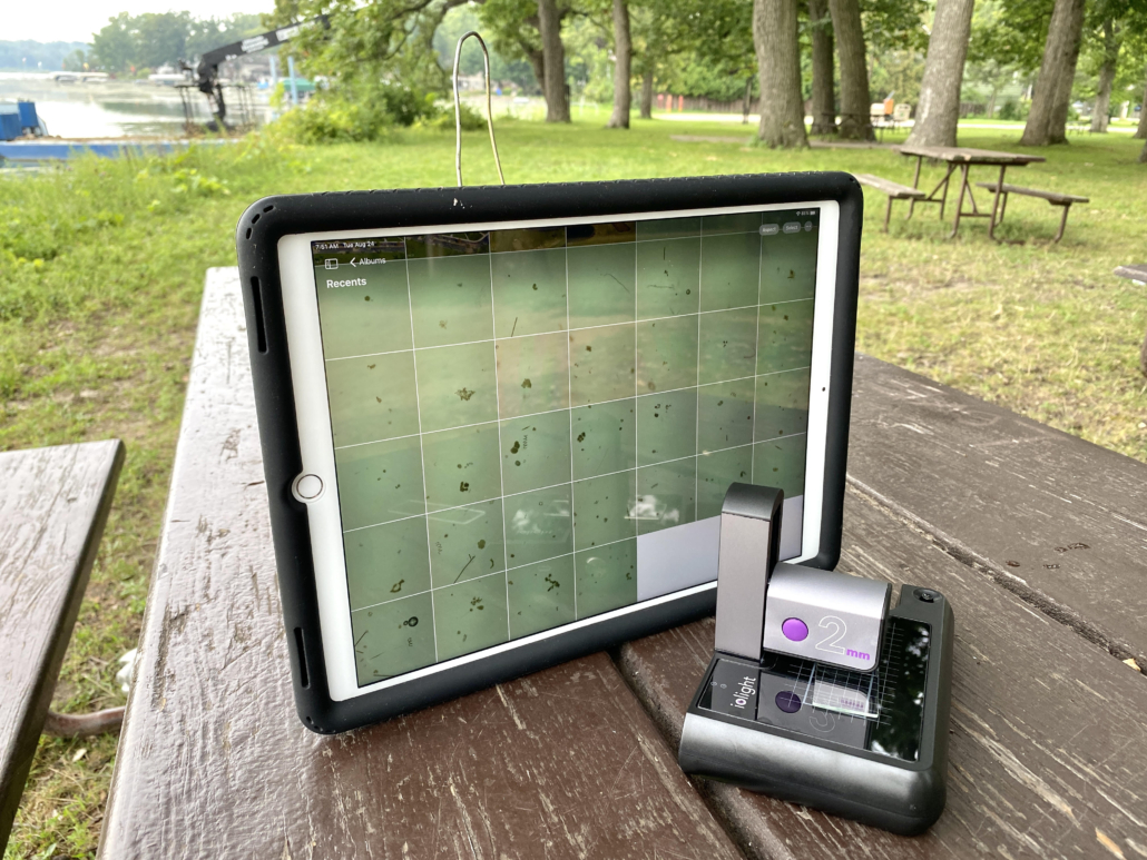
Ramboll spinout, BloomOptix is developing an Artificial Intelligence app to give an instant result on a mobile phone. The expert app is currently being trained using images from lake associations in Wisconsin and New York State. Samples are collected from the lake and placed on the platform of an ioLight pocket microscope and saved onto a mobile phone for analysis. The app will cut the time to diagnose Harmful Algal Blooms from days to minutes. This will maximise the time we can enjoy our beaches and reduce losses to businesses that rely on tourism.
Andrew Monk, CEO of ioLight says “the ioLight microscope is compact and simple for anyone to use, yet still produces high enough quality images for the BloomOptix app to identify toxic algae”. Bryan Arndt, Managing Consultant at Ramboll Water says “The ioLight microscope is twice the quality and half the price of other microscopes we tested.”
ioLight microscope counting algae on a lakeside in the Yahara River watershed, Wisconsin. Image courtesy of Bob Bertra Clean Lake Alliance.
About Ramboll
Ramboll is a global engineering, architecture and consultancy company founded in Denmark in 1945.
Their 16,000 experts create sustainable solutions across Buildings; Transport; Water; Environment & Health; Architecture, Landscape & Urbanism; Energy and Management Consulting.
Across the world, Ramboll combines local experience with a global knowledge base to create sustainable cities and societies. They combine insights with the power to drive positive change to our clients, in the form of ideas that can be realised and implemented.
ioLight has patented the only pocket microscope that can share images of single-cell organisms such as algae and diatoms. ioLight’s solutions allow professional and amateur scientists to measure the effects of climate change such as Harmful Algal Blooms.
ioLight’s customers have taken their microscopes to The Arctic, Antarctic, The Amazon and even up Mount Everest where conventional microscopes cannot be used.
ioLight’s Magnificent Mobile Microscope goes wherever researchers go.
Cells grown in labs are a mainstay of biomedical research. Scientists have available to them a wide variety of carefully selected cells, cultivated from animals and humans. These cells can be grown in the lab so that scientists can test potential therapies, drugs and vaccines on appropriately selected cells to see the effect and then infer what might be the effect when used on live humans and animals.
A cell line is created by harvesting some cells from an animal or human and growing them in flasks. As the cells grow they multiply, and so by continually cultivating the cells, there is a constant supply of new cells identical to the original cells.
There are a large variety of cell lines for various parts of many animals. Each is suitable for different types of experiments and for investigating different biological processes. For example, the CHO-K1 cell line is epithelial cells that come from a particular Chinese Hamster’s Ovary (hence CHO) and is commonly used to test protein therapeutics. Calu-3 is another example of a cell line – this was originally taken from an individual human lung cancer, and so when it is used in the lab, it is often used to investigate and test cancer therapies.
Cells are widely used across a variety of biomedical research topics, but regardless of the research topic, the cell culture techniques required are very similar. A whole industry has grown up to provide cell culture techniques and equipment.
Cells need very carefully controlled conditions to grow successfully, to provide these conditions an incubator is used. The incubator promotes cell growth by maintaining the optimum temperature, humidity, and often atmosphere. Cells typically take between many hours and days to grow, and for the effects of the experiments to be visible. Thus the cells need to be imaged regularly over the course of hours/days both to determine a) that the cells are growing as expected and b) to see the effect of the experiment on the cells. Conventionally this is done by removing the flask from the incubator, putting it on a microscope and looking at the cells. However moving the flask and taking it out of the controlled incubator environment adversely affects the cells, so images are recorded infrequently to minimise the disruption to the cells.
What is Live Cell Imaging?
Live Cell Imaging refers to recording microscope images of the cells without moving the cell culture flask or removing it from the incubator. This minimises failed experiments and enables the scientist to better track the effect of the experiment on the cells. This improves the quality of the data and gives a better understanding of the experiment.
Conventional microscopes are large, complex and cannot be used inside incubators. Instead, micro-incubator chambers that fit on microscope stages are used. However, this approach is expensive and ties up a valuable bench microscope for the entire duration of the cell culture, which makes this approach impractical for most research projects.
Recently, miniature digital inverted microscopes have become available. These microscopes image cell flasks, dishes or well plates from below and record images whenever the researcher wants without disturbing the cells. They are small enough to fit inside standard incubators, making Live Cell Imaging accessible to a much wider range of budgets.
Many of these inverted incubator microscopes are supplied with sophisticated software to automatically track cell development and provide the researcher with high-level data on the progress of cell growth. This is appropriate for many large scale development labs. However, much cutting-edge research is done by the smaller labs for whom this level of sophistication and cost is not appropriate.
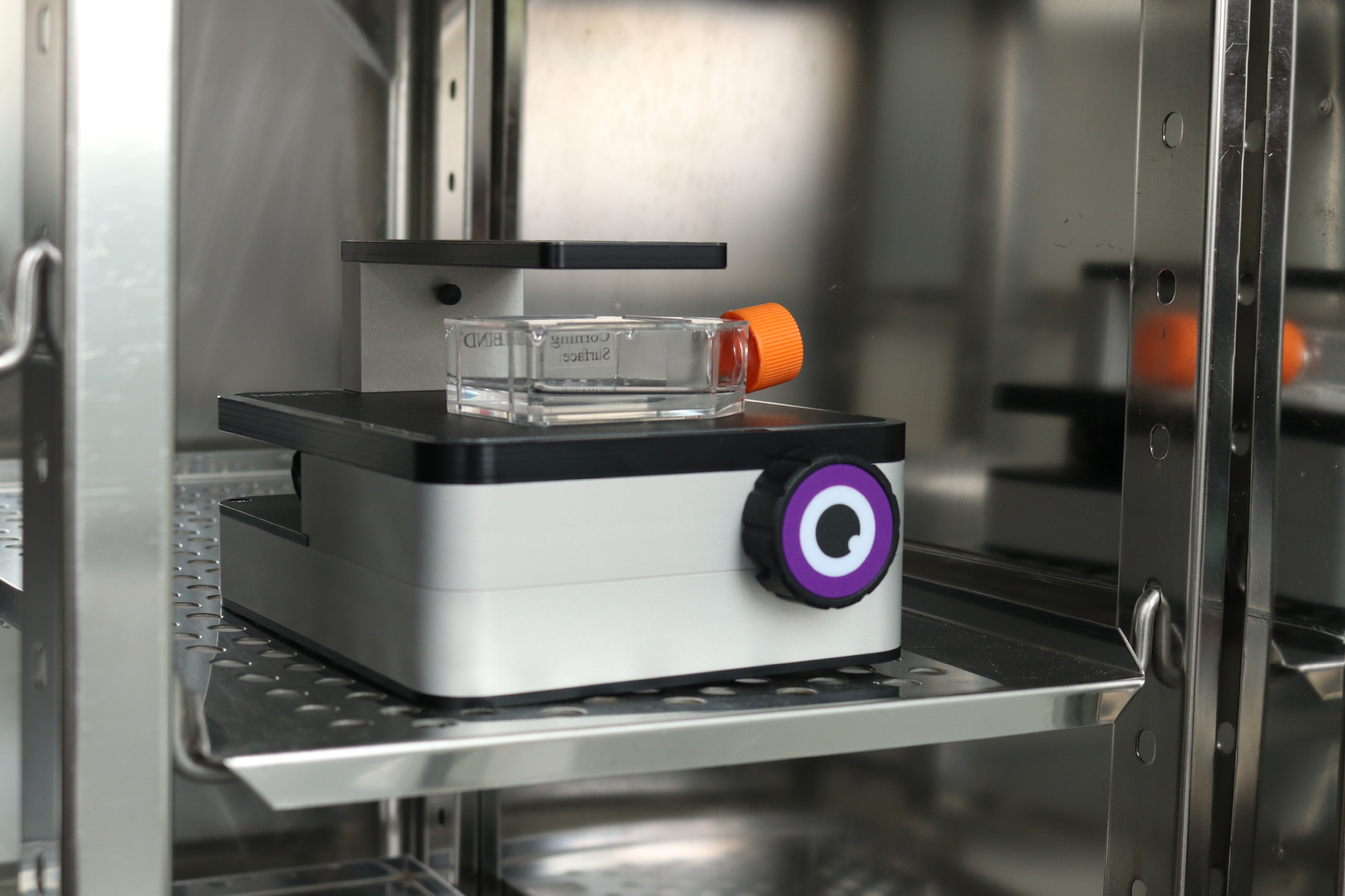
Above: Inverted digital microscope inside an incubator
The images below are taken using a simple low-cost inverted incubator microscope made by ioLight (iolight.co.uk). This microscope fits inside standard incubators and sends images via WiFi to a tablet or computer in the lab. It can record time-lapse images which can be viewed remotely, so the researcher can monitor cells for days without going into the lab or removing the culture from the incubator. This reduces the number of failed cell cultures and provides better data on the results of the experiments.
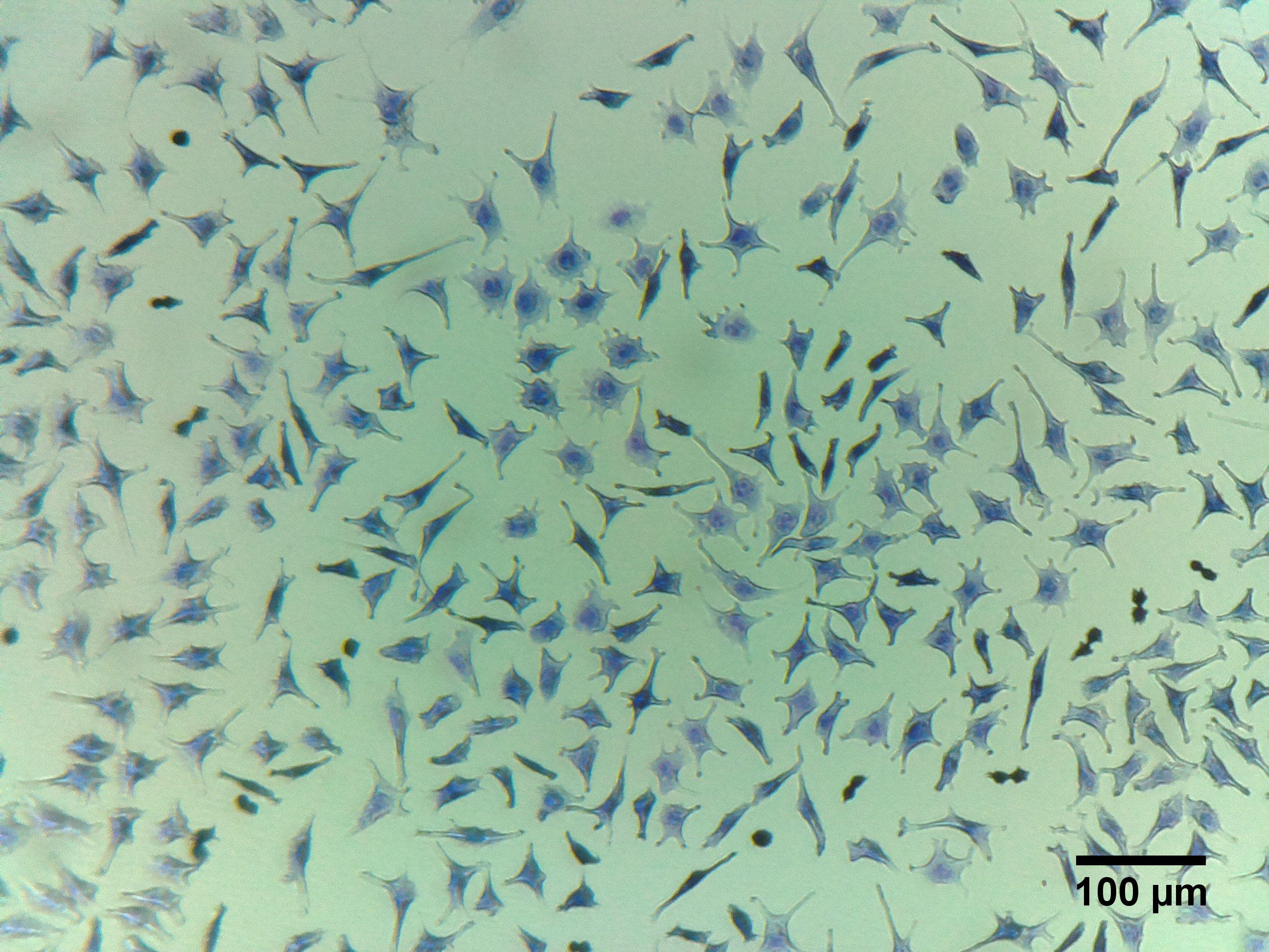
This type of affordable digital inverted microscope is suitable for imaging a wide variety of cells as well as a myriad of other samples. Some example images of cells are shown below:
Live Cell Imaging: Hepatocytes
Hepatocyte cells make up the majority of the cells of a liver and are involved in a range of functions including protein synthesis and storage and transformation of carbohydrates and formation of bile.
Fibroblasts are cells that synthesize the structural framework for animal tissues. They are the most common cells of animal connective tissue. They are used in a variety of applications including stem cell research, and vaccine development.
These cells are cloned from bone marrow and are particular types of neuronal cells often used to study Parkinson’s disease, neurogenesis and other characteristics of brain cells.
Live Cell Imaging is the best way of ensuring a cell culture is progressing correctly and reduces the time and money wasted on failed cell cultures. Live Cell Imaging also enables the researcher to record better data on the effect of the experiment on the cells, thus improving the quality of research. Until recently Live Cell Imaging has been very expensive and the preserve of large labs. However, simpler Live Cell Imaging devices such as the ioLight inverted microscope are now available. Smaller labs, which are often where much of the cutting edge research is done, can use these new microscopes to access many of the benefits of more costly Live Cell Imagers.
https://iolight.co.uk/wp-content/uploads/2021/09/IMG_8614-scaled.jpg13332000ioLighthttp://iolight.co.uk/wp-content/uploads/2016/01/iolight-logo.pngioLight2021-09-02 16:08:192021-09-06 14:35:58Live Cell Imaging on a budget







 ioLight Limited 2024