
Rapid, in-field Stomata imaging with ioLight
Counting stomata and observing their physiology tells us much about Plant growth and productivity. These microscopic pores are key for plant photosynthesis but also play an important role in water loss from the plant. Furthermore, certain pathogens use these pores to access the plant, with the size of the stomata playing a limiting factor. Climate change is creating new challenges to important agricultural crops with increased temperatures, droughts and a growing number of crop pathogens. Developing a balance between drought resistance, yield and susceptibility to new pathogens is becoming increasingly key in the development of new crop strains to face the challenge of changing biomes.
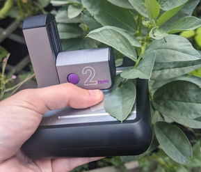
Rapid phenotyping of stomata could be a key tool in creating resilient crop strains. However, current methods are ill-suited to high throughput screening in the field. Collecting and creating a mounting of prospective plant leaves is a labour-intensive and time-consuming task, often making such research prohibitive. ioLight microscopes can image stomata on the leaf directly and in the field. Images can be generated rapidly for counting and analysis by humans or AI.
Using the ioLight microscope


The images above were taken using the ioLight 1mm and 2mm folding microscopes. They show the stomata of a broad bean leaf (Vicia faba). The leaf remained attached to the stem and was flattened using a glass slide to ease imaging, as shown in the picture at the top of this article. Using the ioLight microscope’s high contrast settings, stomata and their surrounding guard cells can be discerned, with the 1mm field of view offering greater detail, whilst the 2 mm field of view microscope offers a greater sampling area.
ioLight microscopes can be held in one hand and imaging takes seconds. Multiple points on the same leaf or multiple leaves on the same plant can be imaged to improve the reliability of results. This rapid imaging speed allows a greater number of plants to be imaged and makes stomatal counts suitable for high throughput screening of factors affecting crop resilience and yield.
Automated Stomata counting
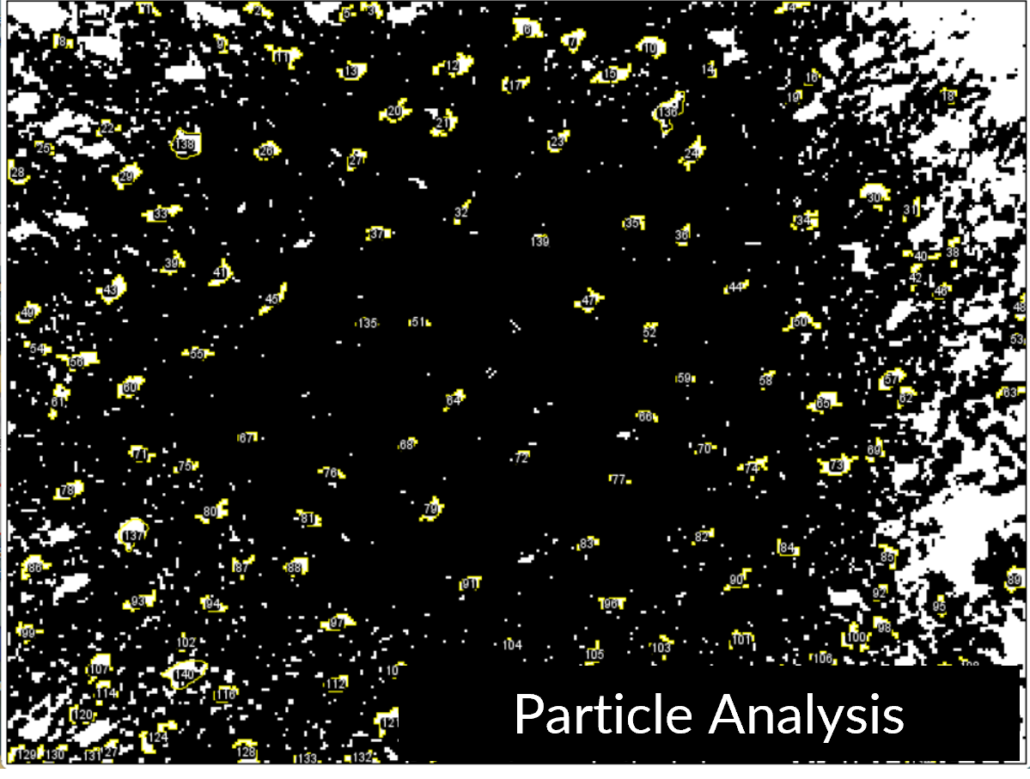
The ioLight microscope produces a digital image on any connected phone, tablet or laptop. This image is readily available for further analysis. In this case, free 3rd party software, imageJ, was used to count the 140 stomata present in the image. Currently, this process involves manual steps, such as thresholding the image to highlight the stomata further and setting the measurements of circumference and circularity for the particle analyser. Nevertheless, the method is faster than a manual count. Further improvements in image processing time can gained by using AI to carry out stomatal counts and gain more insight into stomatal size and state.
Conclusion
Stomata imaging allows scientists to understand further the interplay between the number of stomata and their control in crop species facing the challenges of environmental stress as a result of climate change. Until recently, the ability to capture images of stomata has been a time-consuming and labour-intensive process. However, ioLight microscopes can image stomata directly in the field, unlocking the potential of rapid, high throughput stomatal imaging.

 ioLight Limited 2024


The ioLight microscope offers a revolutionary approach to rapid, in-field stomata imaging, making it easier to analyze plant resilience and productivity under changing environmental conditions, offering significant potential for agricultural research and crop development. Regard Tel U
Thank you for your comment
i want you to send me the price and quotation of Stomata Counter Microscopic image based fully automated stomata detector and pour measurement method
Thank you. We will Email a quotation